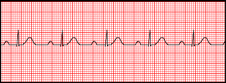
Heart Blocks Can occur in the SA node, AV Node, or in larger sections of the ventricular condition system
Types of Heart Blocks:
1. Sinus Block
2. AV Blocks
3. Bundle Branch Blocks
4. Hemiblocks
Sinus Block-an unhealthy SA node quits pacing activity for at least one compete cycle. The P waves before and after the block are identical because the same SA node pacemaker is functioning before and after the pause.
AV Blocks-when minimal delays the impulse within the AV node, making a longer than normal pulse before stimulating the ventricles.
-Types of AV Blocks
1. First Degree AV Block
2. Second Degree AV Block Type I
3. Second Degree AV Block Type II
4. Third Degree AV Block
First Degree AV Block-prolonged PR interval greater than 0.2 seconds. There is no dropped QRS complex
Second Degree Type I AV Block-the PR interval become lengthened until the P wave does not elicit a responding QRS complex. The PR interval is greater than 0.2 seconds. Also called Wenckebach
Second Degree Type II AV Block-also called Mobitz II is when an occasional ventricular depolarization (QRS complex) is not conducted but dropped. The P waves are normal and normal PR intervals are present.
Third Degree AV Block-also called complete heart block. This is when none of the atrial depolarizations conduct to the ventricles. The P wave rate and the QRS complex rate are independent and will march out with calipers uniformly.
Bundle Branch Blocks-
-Normally the right and left bundles transmit the electrical change to the right and left ventricles at the same time.
-When there is a bundle branch block, there is a delay in the depolarization stimulus that creates the widened QRS complex in the classic rabbit ear pattern (R and R' waves)
-If there is a bundle branch block, look at the chest leads right (V1 and V2) and left (V5 and V6)
Right Bundle Branch Block
-If there is a R,R' waves in V1 or V2 there is a right bundle branch block
-If it is only present in one of the leads it is an incomplete right bundle branch block
Left Bundle Branch Block
-Sometimes the R R' waves will only be seen as notch in the wide QRS complex in V5 and V6
-In a left bundle branch block the left ventricle fires late so the first portion of the QRS complex is right ventricle activity. We cannot see Q waves originating from the left ventricle because they are buried in the QRS complex
Right Bundle Branch EKG:
Left Bundle Branch EKG:
Wolff Parkinson White:
-There is an accessory pathway for ventricular stimulation causing a delay in ventricle stimulation
-This occurs between the SA node and AV junction
-This produces a delta wave
-These patients are prone to SVT
Delta Wave:
Hemiblocks:
-These are blocks of the anterior and posterior division of the left bundle branch
-These are mostly due to loss of blood supply to the anterior or posterior branches of the left bundle branch


No comments:
Post a Comment